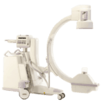
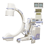
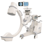
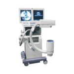
An estimated 4-10 million interventional pain procedures are performed annually in the United States, where at least 50% of those are performed under fluoroscopy. The major purpose of fluoroscopy is for correct needle placement to ensure target specificity and accurate delivery of the injected drug. However, this interventional technique of fluoroscopic guidance does result in radiation exposure with risks posed to patients, physicians, and other personnel. Federal regulations do limit the maximum output for C-Arms, and there are a range of techniques to reduce the risk in private practice settings.
Radiation Exposure
Fear over patient radiation doses is a valid concern. Nonetheless, it is important to understand that the goal of all interventional radiology procedures is to treat (not harm) patients and improve their well-being. As a physician, radiation risks associated with interventional procedures should be discussed with patients, particularly when the expected dose of radiation may be high.
In general, fluoroscopy delivers a dose of approximately 5 rads per minute in the direct beam. The thickness of the patient also determines the exposure rate: the thicker the patient, the higher the exposure. Even small doses of radiation (1 rad = approximately one chance in 100,000) can damage DNA, which can essentially cause acute health effects.
Since shielding the patient is not usually possible, time and distance are key in helping reducing their exposure. Time: shorter fluoro times can be achieved when the physician uses intermittent fluoroscopy (as opposed to continuous), and utilizes the image hold capacity. Distance: the patient’s exposure increases exponentially the closer he/she is positioned to the x-ray tube. Positioning the patient as far as possible from the x-ray tube (maybe 12 to 15 inches away from body) and as close to possible to the image intensifier can reduce exposure. (Decreasing x-ray field size can also reduce patient exposure).
With C-Arms, education is a must for properly monitoring and reducing exposure levels for patients and staff. Receive training from a qualified expert (radiologist or radiological health physicist) in safety procedures and proper imaging techniques. Good safety practices, such as the ones below, can also keep radiation doses As Low As Reasonably Achievable (ALARA).
Radiation Safety
Time, distance, and shielding are the three basic guides that could and should be taken for radiation safety. First and foremost, do not allow any unauthorized visitors during x-ray exams. Only individuals required for the radiographic procedure should be present in the radiographic room during exposures.
Timing. As we mentioned before, with timing, shorter fluoro times can be achieved when the physician uses intermittent fluoroscopy. Also, analyze original radiographs before performing a fluoroscopic examination. Viewing original radiographs, especially for orthopedic studies, can dramatically reduce the repeat rate for the time required for procedures.
Distance. Observers should stand on the image intensifier side of the C-arm if possible, to avoid radiation leakage from the x-ray tube. When not assisting, step away from the patient during fluoro, as feasible. Stepping even one foot further back can significantly reduce a person’s dose.
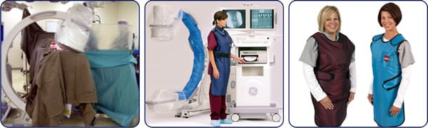
Shielding. In interventional fluoroscopy procedures the tissue of concern is the skin. The skin that is at the site where radiation enters the body receives the highest dose than any other body tissue. All workers in the x-ray room during studies must have a lead apron, and other appropriate shielding wear.
- Lead Apron and Thyroid Shields. Verify that the apron is 0.5 mm lead equivalent, and be sure aprons and shields are in good condition (remove any damaged ones). Insist on well fitting protective gear with a weight your body can handle. Hang aprons and shields on racks. Do not bend or fold them as this can cause cracks and tears in the protective material, making exposed body parts susceptible to radiation.
- Wrap-around Apron. When wearing lead aprons, it is imperative to keep the lead between you and the x-ray tube. Meaning, do not turn your unshielded back to the x-ray tube. If you do need to move about the room where exposing your back may be likely, insist on using a wrap around style apron.
- Leaded Eyeglasses. Those who routinely fluoro for long or interventional procedures, lead glasses with side shields can provide additional protection to the lens of the eyes. Remember, you may need to look sideways from the C-arm x-ray tube to see the image on the monitor, which will leave the lens unprotected if glasses do not have side shields.
- Leaded Gloves. Lead gloves are required if hands are potentially close to or in the primary beam.
Get Started
Request Pricing Today!
We’re here to help! Simply fill out the form to tell us a bit about your project. We’ll contact you to set up a conversation so we can discuss how we can best meet your needs. Thank you for considering us!
Great support & services
Save time and energy
Peace of mind
Risk reduction
Additional Safety Tips
- For needles, try using just one hand so you can keep your other hand away from the needle, and any potential exposure. Be sure there is ample lighting for accuracy. Properly dispose of used needles immediately after use.
- If possible, structural shielding (such as ceiling-mounted lead acrylic shield and an under-the-table shield) should be implemented.
- Always check for damages. If the c-arm or fittings are damaged, the X-ray tube and intensifier may become misaligned, resulting in image degradation or loss, as well as presenting a potential injury to the staff and patient.
- The fluoroscopy beam-on time and x-ray field size should be reduced as much as possible, and the x-ray beam kept well collimated. Failure of the x-ray beam collimation may lead to primary beam x-ray exposure outside of the selected image intensifier input area. This would result in image degradation and additional exposure for the patient.
- Pulsed fluoroscopy, single pulse fluoroscopy mode, manual mode, fluoroscopy timer warning, and last image hold (“freezing the screen”) are also good safety practices.
There are ways radiation exposure can be fine-tuned to help lower radiation levels. Those who use radiology equipment must be adequately trained in equipment operation and radiation safety principles to protect the patients and personnel that are subject to exposure.
Give me a call or drop me a line if you have additional questions or safety tips to share.
Posted by:
John Brant (JB)
Sales Manager
407-438-7847
John@amberusa.com