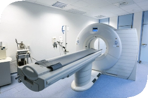
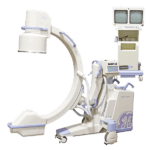
A mammography sounds like any other diagnostic imaging procedure. When you think about it you think of a dark room, a loud machine, and someone in a white coat telling you it’s going to be okay. It’s fair to ask what sort steps take place before, during and after the procedure and what are some of the risks.
So let’s start with what’s known:
- It’s an X- Ray of the breast
- It helps detect tumors on the breast, if they are there.
- Finds abnormal areas and proves whether or not they are cancer
- The procedure has been used for over 30 years
Those are just some fairly common facts about mammography. The procedure itself is a little bit more complicated and requires more of an in depth explanation. First, there are two different types of procedures that are done and they are:
- Screening mammography
- Diagnostic mammography
A screening mammography is done in order to detect changes in the breasts of women that are not showing any common signs of cancer. The procedure involves two X- Rays of each breast. This particular test has the ability to detect a tumor that’s not felt. If a woman doesn’t feel a lump it doesn’t mean that she does not have something to worry about.
A diagnostic mammography checks abnormalities found in a screening mammography. A big part of the diagnostic mammography deals with abnormalities that include pain, thickening of the nipples, undetected masses, even discharge. There is a difference in between the two is fairly easy to see.
A screening mammography works for the purpose of making sure that there’s nothing wrong. The diagnostic mammography sees what’s wrong and goes from there. The two can occasionally work off one another although it’s not necessarily the case. It’s important for the patient to be informed as to what the types of mammography. It’s vital for the physician to understand and properly explain the procedure from the before procedure protocol to the actual procedure itself.
Given the strong advocacy for breast cancer, most women understand the need for a mammography. If a woman is older than 25 and feels lumps, or some sort of unusual symptom, it’s important to get that checked out. If a woman has first degree history she may be well served to get checked out often.
A before the procedure list is always important and it should read like this:
- A thorough explanation that will include answering any questions from the patient
- Signing required forms such as consent.
- Notifying the doctor of pregnancy, implants, breastfeeding or any general developments.
- Understanding do’s and don’ts such as proper attire, avoiding use of powders, perfumes and ointments.
- Ensuring the exam is scheduled for two weeks after the start of menstruation.
- Any specifics stemming from concerns with regards to patient health.
The procedure itself is like any other diagnostic imaging procedure in that it has a list of sorts that’s followed by the person performing it. Once the patient is in, the usual protocol calls for the removal of anything that may interfere with the procedure. The following is a list of what’s done:
- Remove all clothing from the waist up.
- Get the reliable hospital gown to wear.
- The patient will be asked to stand in front of a mammography machine and one breast will be placed on the X-ray plate. In order to position the breast for optimal imaging, the technologist may examine and/or palpate the breast before placing it on the plate. An adhesive marker may be applied to any moles, scars, or other spots that might interfere with the breast image.
- A separate flat plate is brought down in order to gently compress the breast against the X-Ray plate.
- The patient is asked to hold their breath during the procedure.
- Two pictures of each breast are taken at different angles.
- After the pictures are taken, the radiologist examines the film while asking you to wait.
The procedure is known as slightly uncomfortable due to how the breast is manipulated in order to take the picture itself. After a 30 to 45 minute wait the patient will know what the next steps are, if there’s something to worry about or not. The mammography has its share of risks as does any diagnostic imaging procedures. The risks are relatively minor but they are important to acknowledge and they include:
- Radiation Concerns
- Discomfort from breast manipulation
- Interpretation difficulty due to young age
- Interference due to talcum powders, hormones, breast implants, previous surgery.
After a mammography a patient may be on the way home with great news or further instructions. Usually the latter means that there is something to be concerned about. The procedure is a major aide and one way or another it is imperative to have it done.
If you have any questions about mammography systems or any other diagnostic imaging systems please feel free to give us a call. Our team of dedicated professionals here at Amber Diagnostics looks forward to answering any questions you may have.
Bobby Serros
President/CEO
407.438.7847
bobbys@amberusa.com
References: