Skip to content
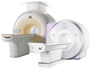
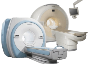
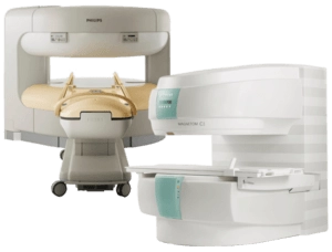
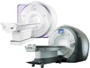
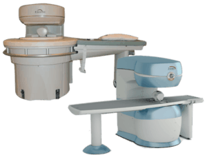
Amber Diagnostics has an extensive selection of high-quality fully refurbished used MRI machines for sale at competitive prices
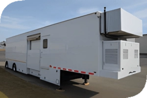
Mobile Options Mobile MRI Machines Our top-tier mobile MRI machines are available to ensure seamless patient care and uninterrupted diagnostics.
For Rental For Sale Featured Our Most Requested Choose The Best Why Amber Diagnostics MRI Machines? Since 1994 Amber Diagnostics has offered a wide array of quality refurbished and used MRI scan machines for sale. In our 25,000 square foot warehouse we offer end to end service for every preowned equipment we house. We have installed MRI scan machines for urgent care facilities, outpatient departments, private practices, and major medical centers throughout the world.
During your consultation, our equipment experts will discuss your department’s needs, budget, and space requirements. We will support you in every stage of the process; from site planning to shipping, installation and technical servicing. Keep your costs down and imaging capabilities high with Amber Diagnostics.
Contact Us Today Close Contrast
Choose color black white green blue red orange yellow navi
used mri machine for sale, siemens, magnet, philips, patient, radiology, hitachi, medical imaging, magnetic field, warranty, toshiba, medical device, brand, esaote, inventory, inspection, cost, gradient, unit, equipment, disease, resonance, manufacturing, technology, general electric, price, machine, software, helium, aperture, slew rate, amplitude, health care, radio, image, ultrasound, contract, tissue, injury, brain, gadolinium, radio wave, claustrophobia, contrast agent, information, blood vessel, metal, blood, intercom, health, liver, radiation, spinal cord, physician, accessibility, noise, exam, advertising, pregnancy, medication, mri contrast agent, organ, bone, neuroimaging, headache, signal, breast, atom, pain, tomography, allergy, abdomen, ionizing radiation, inflammation, insurance, hearing aid, computer, soft tissue, radiography, magnetic resonance imaging of the brain, nerve, research, proton, water, positron emission tomography, data, hydrogen atom, hydrogen, artificial intelligence, injection, therapy, headphones, food and drug administration, mri scan machine price, mri scan machine cost, molecule, uterus, jewellery, anesthesia, joint, physics of magnetic resonance imaging, anatomy, muscle, deep learning, communication, cell, prostate cancer, molecular imaging, medicine, artery, malignancy, knee, dementia, wave, nursing, pelvis, clothing, diagnosis, bone tumor, edema, productivity, iterative reconstruction, sedative, pulse, fluid, kidney disease, pump, neurology, dye, cerebrospinal fluid, cardiology, arteriovenous malformation, workflow, tool, osteomyelitis, surgery, raymond damadian, electric current, nuclear medicine, glasses, drug, health care provider, seizure, learning, lung, back pain, central nervous system, medical history, aorta, button, skin, risk, human brain, oxygen, diffusion, innovation, tractography, energy, education, acceleration, arm, fear, biliary tract, science, ankylosing spondylitis, clinic, perfusion, hives, ear, cardiac imaging, motion, sensitivity and specificity, fibrosis, bile, ulcerative colitis, tendon, shortness of breath, magnetic resonance, mri images, healthcare, brain mri, mri exams, mris, images, mri, mri procedure, mri technologist, mri systems, imaging magnetic resonance, mri scan, mri scans, magnetic, mri technician, diagnostic imaging, imaging, mri machines, medical, mri machine, mri scanners, healthcare provider, siemens healthineers, functional mri, brain imaging, mri exam, clinical, system, mri equipment, open mri scanners near me, mri scan machine, mri scanner cost, mri equipment cost, medical imaging equipment, mri scanner price, mri equipment for sale, neck, statistics, artifact, compressed sensing, ligament, vagus nerve, nausea, echocardiography, nervous system, bladder, ischemia, user experience, intelligence, mri machine for sale, budget, ge healthcare, optima, spain, united kingdom, denmark, netherlands, speed
used mri for sale, mri machine for sale, used mri machine for sale
