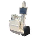
The C-Arm is commonly used for intraoperative imaging and in the fields of orthopedics, traumatology, vascular surgery, and cardiology
You may know the C-Arm as an imaging device that uses X-Ray technology and has a C shape, making it able to be used flexibly in many different areas within a clinic.
The C-Arm is commonly used for intraoperative imaging and in the fields of orthopedics, traumatology, vascular surgery, and cardiology. In the operating room, C-Arms help visualize kidney drainage, abdominal and thoracic aortic aneurysm repair, percataneous valve replacements, cardiac surgery, vascular surgery, gastroenterology, neuro stimulation, pain management, neurology procedures, and more.
The C-Arm comprises of an X-Ray source and an image intensifier or flat-panel detector. The C shape of the machine comes in handy as it allows the system to move any way needed in order to garner images of the patient.
Get Started
Request Pricing Today!
We’re here to help! Simply fill out the form to tell us a bit about your project. We’ll contact you to set up a conversation so we can discuss how we can best meet your needs. Thank you for considering us!
Great support & services
Save time and energy
Peace of mind
Risk reduction
When the X-Rays enter the patient’s body, the image intensifier or detector converts the X-Rays into an image that shows up on the C-Arm monitor. Then the physician present can check anatomical details at any point, allowing surgeons to monitor progress in real time; that way, if any mistakes are made, they can be corrected immediately.
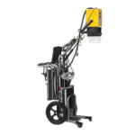
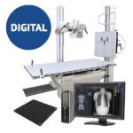
One can choose from an analog C-Arm or a digital C-Arm, analogs using image intensifiers with lower image quality, but available at lower prices; and digital C-Arms, which use a flat panel detector and produces better image quality, but at a higher price point. In addition to Full Size C-Arms there are Mini C-Arms, which are better suited for sports medicine, orthopedic, and podiatric imaging.


