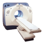
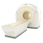
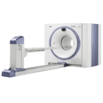
A CT Scanner and a PET/CT scanner are both a type of imaging machines that provide information about internal structures of the body
A CT Scanner and a PET/CT scanner (Positron Emission Tomography-Computed Tomography) are both a type of medical imaging machines that provide detailed information about internal structures of the body.
The PET (Positron Emission Tomography) scanner is a medical imaging device that uses a small amount of radioactive material, called a radiotracer, to create detailed images of the body’s cells and tissues. The radiotracer is injected into the patient’s bloodstream, and then a special camera detects the gamma rays emitted by the radiotracer as it decays, creating an image of the body’s internal structures.
A CT (Computed Tomography) scanner, on the other hand, uses X-rays to create detailed, cross-sectional images of the body. The X-rays are emitted from multiple angles, and a computer then uses the information to create a detailed image of the internal structures of the body.

Get Started
Request Pricing Today!
We’re here to help! Simply fill out the form to tell us a bit about your project. We’ll contact you to set up a conversation so we can discuss how we can best meet your needs. Thank you for considering us!
Great support & services
Save time and energy
Peace of mind
Risk reduction
When used together, a PET/CT scanner combines the detailed information provided by a PET scan with the high-resolution structural information provided by a CT scan. This can help doctors identify the location and function of abnormal cells and tissues, as well as provide detailed information about the anatomy of the affected area. This can be particularly useful in diagnosing and staging certain types of cancer, monitoring the progress of a treatment, and assessing the effectiveness of a treatment.
It is important to note that CT scans are mostly used for diagnostic imaging, where as PET – PET/CT is used for functional imaging and metabolic information of the body.
PET/CT scanners combine the capabilities of both machines, providing both functional and structural information about the body. However, keep in mind that CT scan has a higher exposure to ionizing radiation and PET scans involves the use of radioactive tracers. Therefore, it is important to consider these and other factors when determining which machine is best for a specific patient.


