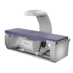
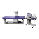
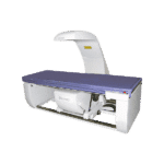
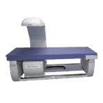
The Hologic Horizon Wi bone density machine is the most rapid means of acquiring accurate BMD results

Hologic Horizon Wi Overview
The Horizon DXA System enables you to capture highly detailed images you can trust – even when imaging large, obese patients. With its low-noise detectors, you have the power to assess an expanded range of clinical conditions with speed and precision.
Hologic’s detector array also works in concert with a new high-capacity X-ray generator to increase heat-load capacity for longer life — with no cool-down time. Not only does this help increase patient throughput, it improves image quality.
Benefits:
Femur Fracture Assessment: Horizon DXA produces radiographic quality images of the entire femur for assessment of potential atypical femur fractures. A quick, 15-second scan reveals cortical thickening of the bone, making it fast and easy to monitor the effects of bisphosphonate therapy over time.
Abdominal Aortic Calcification: Visualize calcified plaque in the abdominal aorta, which may be a significant indication of heart disease and stroke – two of the leading causes of death in men and women.
Instant Vertebral Assessment™ Scan: Assess fracture risk by combining an accurate measurement of bone density with high-resolution vertebral imaging. You can identify spine fractures with a low-dose, single-energy image in 10 seconds.
BMD Histogram: Improve accuracy and reduce post-exam analysis errors with precise, software-assisted placement of inter-vertebral disc spaces for graphic analysis.
Internal Dynamic Calibration System: Hologic’s exclusive Dynamic Calibration System delivers pixel-by-pixel calibration through bone and tissue equivalents — for greater long-term precision.
OnePass™ Technology: A new digital high-resolution ceramic detector array is paired with true fan-beam acquisition geometry to enable rapid, dual-energy bone density measurements in a single-sweep scan. OnePass eliminates beam overlap errors and image distortion found in rectilinear acquisition techniques — for superior image quality and data stability. Another Hologic exclusive.
Features:
Exceptional Precision: A whole body composition scan takes as little as three minutes — efficiency without sacrificing accuracy:
- Reflection feature — designed to eliminate the
need for multiple scans, even if portions of the
body lie outside of the scan field - Hologic’s X-ray penetration produces superb
image quality for all patients, regardless of their
shape or size
Enhanced Measurements: Fat Mass Index (FMI) is an obesity classification which measures the ratio of fat mass to height squared.5
FMI may be better than Body Mass Index (BMI), because it is a non-specific measure of excess weight that may misclassify muscular subjects as overweight or obese, interfering with diagnosis’ and management of clinical obesity. FMI is exclusive to Hologic.
Fat Mass Index
- Fat mass ratio not based upon weight (Fat/Height)
- FMI is expressed in units of kg/m2
- NHANES reference — acquired exclusively on
Hologic fan beam systems - Gender specific
- Not affected by lean mass like BMI or %fat
Improved Patient Management and Care: Proper analysis is essential for accurate diagnostic scores making it easier to determine the appropriate course of action for your patients’ management.
The Rate of Change report simplifies patient follow-up by providing comprehensive trending as well as serial tissue mapping. Only Hologic provides an illustration of a patient’s progress using color coded images from previous scans, making it easy for patients and their physicians to track long-term changes.
Measurement of Visceral Adipose Tissue

InnerCore™ Visceral Adipose Tissue (VAT) Assessment:
Deep visceral fat is metabolically active and is often associated with diabetes mellitus, dyslipidemia, hypertension, impaired fasting glucose, impaired glucose tolerance and metabolic syndrome. With Hologic’s InnerCore™ Visceral Fat Assessment, you now have a convenient way to estimate a patient’s visceral fat in the abdominal region, allowing clinicians and researchers a unique understanding of these potential disease processes that may place certain patients within a higher risk category.
DXA accurately and precisely measures whole body, regional fat and lean tissue within the body. Producing consistent results with regard to clinical concerns relevant to obesity related diseases.
- Quantifies total body and regional fat mass with resulting indices.
- Visceral fat area results from DXA correlate with visceral fat area
in CT at L4/L5.5 - VAT Assessment and body composition in one quick whole body scan
- Results are just a few clicks away


