
Fuji has a hand in the medical industry as well, being a manufacturer of computed radiography (CR) machines
Fujifilm, or simply Fuji, is a Japanese film company founded in 1934. Even though they are known mostly for their domestic film and cameras, Fuji has a hand in the medical industry as well, being a manufacturer of computed radiography (CR) machines we sell here at Amber Diagnostics. Read on to learn more about the Tokyo-based conglomerate!
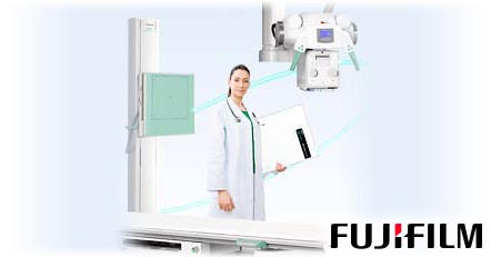
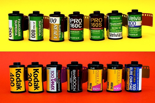
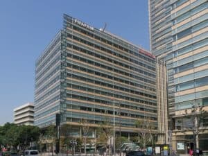
- Fuji was established as Fuji Photo Film Co., LTD, and was based on a government plan to establish a photographic film manufacturing industry.
- Fuji was a major player in motion picture film, however, the company stopped producing the film in 2013 due to the motion picture industry diving more into the digital era.
- In 2006, Fuji released functional skin care cosmetics, something that may seem odd for a film company. The skin care line uses core technologies developed by the research of photosensitive materials.
Get Started
Request Pricing Today!
We’re here to help! Simply fill out the form to tell us a bit about your project. We’ll contact you to set up a conversation so we can discuss how we can best meet your needs. Thank you for considering us!
Great support & services
Save time and energy
Peace of mind
Risk reduction
- Fuji jumped into the medical market in 2003, with the Digital Diagnostic X-Ray Imaging System FCR Profect CS. They later went on to produce Digital Mammography systems and Endoscopy Systems ,as well as CR machines.
- Fuji and film producer Kodak have been in a rivalry for years. Fuji gained a great advantage in the U.S. in 1984, however, during the Los Angeles Olympics: Kodak passed on the opportunity to become the official film of the games, so Fuji won sponsorship rights.
Featured
Our Most Requested
X-Ray Machines
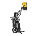
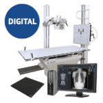
Current Context for CR and DR Buyers
That history is worth knowing. The buying question is simpler: computed radiography and digital radiography are two different workflows, and the difference shows up every day in your department, not in a spec sheet.
CR captures the image on a reusable imaging plate held in a cassette. A technologist carries the plate to a reader, the reader scans it, and the image reaches the workstation from there. DR captures the image on a digital detector and the image moves straight into the workstation workflow. One has a handling step in the middle. The other does not.
Practical Comparison
| Workflow question | CR | DR |
|---|---|---|
| Image capture | Reusable imaging plate in a cassette | Digital detector |
| Processing step | Separate CR reader | Image appears through the detector and workstation workflow |
| Fit with an existing room | Often works with cassette-based rooms already in place | May require detector, workstation, interface, and room integration review |
| Main due-diligence focus | Plate and reader condition, throughput, service, parts | Detector condition, calibration, batteries and cables, workstation and licenses |
For a deeper side-by-side, see Amber’s computed radiography versus digital radiography guide.
Buying or Keeping a Legacy CR System
Age alone does not retire a system. Support does. Before you buy a used CR setup or decide to keep the one you have, find out:
- whether the exact reader model and its plates are still supportable, and whether replacement plates and parts can be sourced
- what preventive maintenance and calibration records exist, and how recent they are
- whether image quality has been tested, and by whom
- which DICOM functions work with your PACS, and which have been worked around
- what the plan is for cybersecurity patching and workstation compatibility over the life you expect
Throughput deserves its own look. The plate-handling step costs time on every exam, and that cost scales with volume. A department running a handful of studies a day feels it very differently from one running a full schedule.
Most of this applies to any used x-ray equipment purchase, not just CR.
Evaluating a DR Upgrade
A DR conversion is not a detector purchase with a cable attached. Confirm:
- detector size and physical fit with your bucky, table, and wall stand
- generator and interface compatibility, including exposure synchronization
- workstation hardware and software licenses
- DICOM, RIS, and PACS integration, including worklist and routing behavior
- wireless coverage or tethering, plus battery and charger requirements
- service support, parts availability, and acceptance testing
Have a qualified physicist or other appropriate professional review image quality and dose-management procedures as part of acceptance. If you are comparing digital x-ray systems, that review should happen before the final configuration is locked, not after.
Planning the Migration
Inventory what you already have before choosing a detector: generator, bucky and stand interfaces, cassette sizes, reader, workstations, network drops, and PACS and RIS connections. Then decide what you are actually doing. A limited detector conversion and a full room modernization are different projects with different budgets.
Settle the questions nobody volunteers. Who configures exam codes, worklists, image routing, and user access? What happens if integration testing fails, and how do you roll back? Which baseline images and quality-control records do you keep so the clinical and physics teams can judge the finished workflow against agreed acceptance criteria?
Put staff training and a downtime plan in the cutover schedule rather than treating them as follow-up.
Common Question
Is CR automatically obsolete?
Not in every setting. Support, parts, workflow demands, and integration are what make a legacy CR system impractical, and those vary by department. Compare the documented operating condition and the full workflow cost against a supported DR option. Deciding from the install year alone gets it wrong in both directions.
Talk Through an X-Ray Workflow
Send Amber your room configuration, your current detector or reader details, your PACS environment, location, and the timeline you are working toward. Availability and project scope are confirmed separately. To start that conversation, contact Amber.
